

四指马鲅(Eleutheronema tetradactylum),隶属于鲻形目(Mugiliformes)、马鲅亚目(Polynemoidei)、马鲅科(Polynemidae)、四指马鲅属(Eleutheronema),俗称马友鱼、午笋、鲤后等[1],是热带、亚热带和温带海域的中上层鱼类,主要分布在印度—西太平洋、波斯湾、新几内亚、澳大利亚、印度、越南、东南亚和中国海域[2⇓-4]。四指马鲅为广盐性溯河洄游型鱼类,幼鱼多栖息于河口,成鱼则多见于沿海水域的浅泥底,具有生长速度快、肉质鲜美、营养价值高、抗病力强等优点,在水产养殖业中具有非常高的商业价值[5] 。2012年至2015年,区又君等[6]成功完成四指马鲅规模化的全人工繁育技术研究,并在沿海地区进行推广,使之成为我国海水鱼类养殖的重要品种。
溶解氧(Dissolved oxygen,DO)是重要的水环境因子之一,对鱼类的生长和存活具有重要的影响。当处于低氧环境下,鱼类自身的生理和生化状态会发生改变,体现在生长、发育、繁殖、行为代谢、抗氧化等多个方面。而为了应对水中溶解氧的变化,鱼类也在逐步适应并做出各种反应及建立机制,通过一系列的生理和行为变化来适应氧环境的变化。
低氧可使鱼体代谢速率降低、摄食能力受到限制、消化吸收率下降等,从而影响鱼体的生长和抗逆性[7⇓-9];鱼体内分泌功能紊乱,从而导致鱼体在产卵期的受精卵品质下降;幼鱼成活率降低,两性比例失衡[10]。目前,低氧胁迫逐渐成为鱼类生理学研究的热点之一。从有关低氧胁迫对鱼类影响的研究发现,低氧水环境会影响鱼类鳃器官的功能,低氧胁迫后,鲫(Carassius auratus)鳃小片之间的空隙完全被细胞团(ILCM)填充,增加了其与水接触部分的表面积[11-12],而且为清除体内产生的大量活性氧自由基,鲫的血清、肌肉和鳃组织中抗氧化酶活力均升高[13]。鳃呼吸表面积增加现象,同样也出现在鲂鲌杂交种F3和西伯利亚鲟(Acipenser baerii)幼鱼中[14⇓⇓-17]。卵形鲳鲹(Trachinotus ovatus)鳃小片细胞表面舒张和抬升,同时毛细血管中的血细胞数量显著增加[18]。许氏平鲉(Sebastes schlegelii)鳃组织形态出现鳃小片基质肥大、增生、末端膨大和上皮细胞水肿等组织病变情况[19]。同样,随着水中溶解氧含量的降低,虹鳟(Oncorhynchus mykiss)鳃小片肥大、增生、末端膨大的比例增加[20]。水体环境多变,为适应水体的溶解氧浓度变化,不同鱼类建立了不同的适应机制 [21-22]。但目前未见关于低氧对四指马鲅鳃和肝脏组织形态变化影响的研究报道。作为鱼类气体交换的关键部位,在受到环境胁迫后,鱼类鳃器官往往容易受到影响并被损伤[23-24];肝脏作为重要的代谢器官,其结构变化与机体生理状态密切相关。因此,本研究采用组织石蜡切片及扫描电镜技术,观察、比较了低氧胁迫条件下四指马鲅幼鱼鳃适应性的变化特征、肝脏组织损伤情况,以及鳃和肝组织对低氧环境的适应性及其组织结构的变化机制,旨在为四指马鲅的苗种培育和养殖生产提供参考。
1 材料与方法
1.1 试验材料
试验样品为中国水产科学研究院南海水产研究所——广东省中山科技成果转化基地自繁自育的四指马鲅幼鱼,平均全长为4.31 cm, 体质量为0.79 g。试验开始前,暂养一周,养殖水温为28℃,水体盐度为4。在此过程中,每天换水一次,换水量为暂养水体的1/3;24 h不间断充气增氧,保持水中溶解氧浓度在6 mg/L 以上。每天9:00、18:00 各投喂 1 次,投喂1 h 后及时清除残饵和粪便,试验前停饲1 d。
1.2 试验方法
1.2.1 试验设计
采用YSIProODO光学溶解氧测量仪(使用前用碘量法校正)测定水体的溶解氧浓度。试验设置低氧胁迫、常规培育对照2个试验组,每组3个重复,共6个水槽(规格30 cm×30 cm×45 cm,实际水体27 L)。随机选取体色正常、体格健壮、活力好的四指马鲅幼鱼,每个水槽放置试验幼鱼10尾。低氧胁迫组溶解氧浓度设计参考与四指马鲅同属鲻形目的鲻(Mugil cephalus)的资料:温度对鲻幼鱼的窒息点和窒息时间影响显著,温度越低,窒息点越低,当16℃时,鲻的窒息点最低,为 0.49 mg/L;在 32℃时,鲻的窒息点最高,为 0.75 mg/L[25]。同时参考具有与四指马鲅类似习性的黄鳍鲷的资料:黄鳍鲷幼鱼在23℃时的窒息点为1.81 mg/L,29℃为4.2 mg/L[26]。并在正式试验前,做预备试验,在28℃静水密闭时,观察到四指马鲅在2 mg/L时偶有浮头现象,80 min出现死亡,120 min死亡过半。因此,本试验低氧胁迫组溶解氧浓度取2 mg/L,并且试验期间通过关闭水槽流水、向水槽内充入氮气,使水中氧气快速溢出而降低水体溶解氧浓度。当水体溶解氧浓度为(1.90±0.12) mg/L 时,将四指马鲅幼鱼放入水槽,并用保鲜膜封闭水槽,在试验过程中通过调节氧气进气阀,使水槽内水体溶解氧溶度维持在2.0 mg/L,进行120 min低氧胁迫试验。对照组则不间断充气增氧,保持水中溶解氧在6 mg/L 以上。
1.2.2 样品采集与处理
在试验2 h后,从每个水槽取4尾鱼,逐尾剖取鳃和肝脏,分别放入2 mL离心管中并分别用4%多聚甲醛溶液固定和电镜固定液固定。
组织切片制作:将固定好的样品以流水冲洗12 h,用50%~100%乙醇进行梯度脱水(50%乙醇2 h、70%乙醇4 h、80%乙醇2 h、85%乙醇2 h、95%乙醇45 min×2次、100%乙醇45 min×2次),再用二甲苯进行透明(1/2无水乙醇+1/2二甲苯的混合液1 h、二甲苯15 min×2次),浸蜡(石蜡2 h×2次),用石蜡进行包埋,切片,采用苏木精—伊红(HE)进行染色,中性树脂封片。
透射电镜制备:透射电镜样本在电镜固定液固定后用0.1 mol/L磷酸缓冲液PB(pH 7.4)漂洗3次,每次15 min;采用1%锇酸溶液避光、室温固定2 h,0.1 mol/L磷酸缓冲液漂洗3次,每次15 min;30%-50%-70%-80%-95%-100%梯度乙醇溶液逐级脱水,每次20 min;100%丙酮2次,每次15 min,再次脱水;用丙酮和812包埋剂渗透包埋,包埋板60℃聚合48 h;超薄切片机切片;150目方华膜铜网捞片;2%醋酸铀和100%乙醇溶液避光染色,70%乙醇清洗,超纯水清洗;2.6%枸橼酸铅溶液避二氧化碳染色,超纯水清洗;干燥,透射电子显微镜(HT7800)下观察并拍照。
1.3 数据处理
运用Excel和SPSS 26软件对数据进行处理,所得数据用平均值 ± 标准差表示。采用单因素方差分析对溶解氧变化的试验结果进行组间显著性分析,将在不同溶解氧浓度下的数据进行LSD多重比较,以 P<0.05作为差异显著的标准。
2 结果与分析
2.1 低氧胁迫下四指马鲅幼鱼的状态
在溶解氧浓度2 mg/L、低氧胁迫时间120 min的试验过程中,四指马鲅幼鱼状态表现为游动时失去平衡,短暂恢复后又再次失去平衡,偶尔跃出水面,重复上述状态后逐渐在游动时不能保持平衡,侧翻静卧缸底并大口呼吸,鳃盖张合不连续,鱼体基本静止,咧鳃后死亡。而对照组幼鱼无任何应激反应,游动状态正常。
2.2 低氧胁迫下四指马鲅幼鱼鳃组织显微结构的变化
图1和表1为低氧胁迫下四指马鲅幼鱼鳃组织显微结构及鳃小片的变化。对照组四指马鲅幼鱼鳃丝上的鳃小片整体排列紧密,完整对称地分布于鳃丝两侧;椭圆形的线粒体丰富细胞位于鳃小片基部与鳃丝相连之处,着色较深;扁平状上皮细胞规则排列;血细胞均匀分布于鳃丝血窦之中,整体结构表现为正常的生理形态。与对照组相比,低氧组四指马鲅幼鱼鳃组织中鳃丝内血窦收缩,血细胞数量增多且分布不均,血细胞在鳃丝末端堆积而使鳃丝末端膨大;鳃小片部分呈弯曲不规则排列,且长度变长、直径变小,鳃小片之间间隔变大;有鳃丝断裂现象;线粒体丰富细胞体积变小,着色较浅;上皮细胞整体被拉长,部分上皮细胞肿胀凸起,甚至破裂;整体形态偏离正常的生理形态。结果表明,低氧胁迫对四指马鲅幼鱼鳃组织显微结构造成了一定程度的氧化损伤。
图1
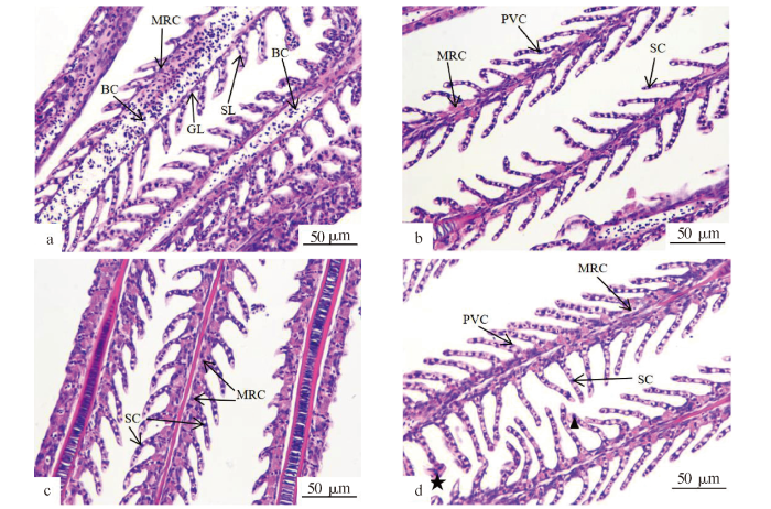
图1
四指马鲅幼鱼鳃显微结构
注:a、c为对照组(400X);b、d为2 mg/L低氧组(400X);GL:鳃丝;SL:鳃小片;BC:血细胞;PVC:扁平上皮细胞;MRC:线粒体丰富细胞;SC:柱细胞;▲:肿胀;★:破裂。
Fig.1
Microstructure of the gills of E. tetradactylum
Notes:a and c were control group(400X);b and d were 2 mg/L dissolved oxygen group(400X);GL.Gill filament;SL.Secondary gill lamella;BC.Blood cell;PVC.Pavement cell;MRC.Mitochondria rich cell;SC.Pillar cell;▲.Swell;★.Rupture.
表1 低氧胁迫下四指马鲅幼鱼鳃小片的变化
Tab.1
| 组别 Group | 溶解氧浓度/ (mg/L) Dissolved oxygen concentration | 鳃小片间隔/μm Space between secondary gill lamella | 鳃小片长度/μm Length of secondary gill lamella | 鳃小片宽度/μm Width of secondary gill lamella |
|---|---|---|---|---|
| 低氧组 Hypoxia group | 2.0 | 18.272±0.41a | 49.288±4.86a | 3.558±0.66a |
| 对照组 Control group | 6.0 | 12.993±1.89b | 31.916±3.54b | 7.071±1.29b |
注:同列数据后不同小写字母表示组间差异显著(P<0.05)。
Note:Different lowercases in the same column indicated significant difference between groups(P<0.05).
2.3 低氧胁迫下四指马鲅幼鱼鳃组织超微结构的变化
图2为四指马鲅幼鱼鳃组织超微结构的变化。在透射电镜下,可以观察到对照组(a、c、e)四指马鲅幼鱼的鳃小片有两层单层呼吸上皮细胞,两层单层呼吸上皮细胞间形成腔隙结构,柱细胞分布在腔隙间并起支持作用,使其形成大片空隙,而形成毛细血管腔,腔内分布有血细胞。鳃小片基部分布有内凹开口的线粒体丰富细胞,细胞内分布大量的线粒体和排泄小泡。低氧组(b、d、f)四指马鲅幼鱼的鳃组织在低氧胁迫前的上皮细胞表面呈褶皱状,胁迫后变平滑;空泡现象加剧;线粒体丰富细胞的顶隐窝由内凹变为平整;线粒体丰富细胞内线粒体形态由椭圆形变为圆形。结果表明,低氧胁迫导致四指马鲅幼鱼鳃组织超微结构中线粒体丰富细胞以及上皮细胞的形态发生变化。
图2
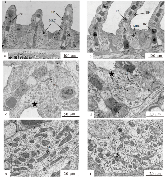
图2
指马鲅幼鱼鳃超微结构(透射电镜)
注:a、c、e为对照组; b、d、f为2 mg/L低氧组;EP.上皮细胞;M.线粒体;MRC.线粒体丰富细胞;BC.血细胞;★.顶隐窝;Pc:柱细胞;N:细胞核。
Fig.2
Ultrastructure of the gills of E. tetradactylum (TEM)
Notes:a,c,e were control group;b,d,f were 2 mg/L dissolved oxygen group;EP.Epithelial cell;M.Mitochondria;MRC.Mitochondria rich cell;BC.Blood cell;★.Top crypt;Pc.Pillar cell;N.Nucleus.
2.4 低氧胁迫下四指马鲅幼鱼肝组织显微结构的变化
图3为低氧胁迫下四指马鲅幼鱼肝组织显微结构的变化。对照组中四指马鲅幼鱼肝组织中,肝小板和肝血窦沿中央静脉呈放射状相间排列;肝细胞多为单核,细胞核大而圆,位于细胞中间且着色较深;细胞之间边界明显,部分出现因糖原溶解而形成的空泡;整体结构清晰,表现为正常形态。与对照组相比,低氧组四指马鲅幼鱼肝组织中肝细胞空泡消失,细胞边界模糊且形状不规则,细胞核变大,细胞胞质减少;细胞排列散乱,细胞核着色较深,部分细胞的细胞核偏离中央、位于一侧;肝小板结构不清晰,肝血窦间隙增大,血细胞数目增加;局部出现肝细胞坏死,肝细胞融合现象;中央静脉的横切面呈不规则形状且内部充满血细胞。结果表明,低氧胁迫对四指马鲅幼鱼肝组织显微结构中肝细胞形态以及血窦分布造成了一定程度的影响。
图3
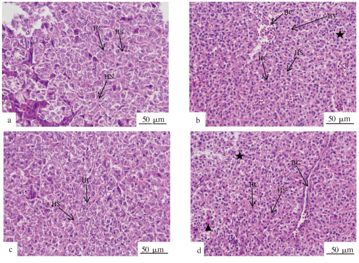
图3
四指马鲅幼鱼肝脏显微结构
注:a、c为对照组(400X);b、d为2 m/L溶氧组(400X);BV:血管;BC:血细胞;HC:肝细胞;HN:肝细胞核;HS:肝血窦;★:肝细胞融合;▲肝细胞坏死。
Fig.3
Microstructure of the liver of E. tetradactylum
Notes:a,c were control group(400X);b,d were 2 mg/L dissolved oxygen group(400X);BV.Blood vessel;BC.Blood cell;HC.Hepatocytes;HN.Hepatocyte nucleus;HS.Hepatic sinusoid;★.Hepatocyte fusion;▲.Hepatocyte necrosis.
3 讨论
3.1 低氧胁迫对四指马鲅幼鱼鳃组织学结构变化的影响
对于大多数水生动物尤其是鱼类而言,鳃是主要的呼吸器官,同时具有摄取食物、排泄氨氮等代谢废弃物、调节渗透压等生理机能。本研究发现,四指马鲅幼鱼在低氧胁迫后,出现鳃组织器官上皮细胞肿胀凸起、抬升以及部分上皮细胞破裂,由扁平状变为梭形及三角形,鳃丝末端充血肿胀,血细胞数量增多,以及血窦收缩等病理现象。这些变化均表明,在2 mg/L溶解氧浓度下的低氧胁迫造成了四指马鲅幼鱼鳃器官组织学损伤,与低氧胁迫下卵形鲳鲹(Trachinotus ovatus)鳃器官上皮出现了肿胀和抬升、鳃小片中血细胞数量增加等试验结果[18]一致。低氧胁迫环境下,四指马鲅鳃器官供氧不足,因而具有运输氧气功能的血细胞数量增多,上皮细胞抬升以增加其与水接触面积,从而获得氧气。此外,低氧胁迫组四指马鲅幼鱼的鳃小片直径变短、长度延长、鳃间隔增加,这与莫桑比克罗非鱼(Oreochromis mossambicus) 在低氧胁迫后鳃丝的长度和鳃小片的面积均增加[27]、鲂鲌杂交种 F3低氧胁迫24 h后鳃小片急剧伸长和鳃小片间距增加[14]等一致。低氧环境下鳃小片需要通过过滤更多的水而获得氧气,而鳃小片的形态变化扩大了其与水体的接触面积,使呼吸表面积增加、水血屏障距离缩短,从而有利于气体交换[28],进而有利于四指马鲅在低氧环境中存活。
3.2 低氧胁迫对四指马鲅幼鱼鳃组织超微结构的影响
上皮细胞与柱细胞及毛细血管之间有一层较厚的基膜,形成特殊的水血屏障双层结构,是鳃丝进行气体交换时隔离内环境和外部水环境的重要结构。有学者认为上皮细胞表面褶皱微嵴,易被黏液覆盖,不利于增加气体交换面积[29],而本研究中四指马鲅上皮细胞表面由褶皱状(对照组)变平滑(低氧胁迫组),这可能是为了增大细胞呼吸面积,提高气体交换率,从而有利于四指马鲅在低氧环境中存活。鱼类鳃器官的调节渗透压以及离子平衡主要由线粒体丰富细胞完成。本研究发现,急性低氧胁迫120 min后,线粒体丰富细胞顶隐窝同样由内凹变为平整,胞内线粒体形态由椭圆形变为圆形,体积增大,与星丽鱼 (Astronotus ocellatus)和虹鳟低氧胁迫后线粒体丰富细胞开口变大 [30]、卵形鲳鲹急性低氧胁迫 6 h后鳃器官线粒体丰富细胞的形态变化及数量增加[18]等结果一致。这可能是在低氧胁迫下,水环境中氧气含量不足,而四指马鲅机体的正常代谢仍然需要相同甚至更多的能量,因此其通过改变线粒体的形态,以更加充分地利用氧气。研究结果表明,四指马鲅幼鱼为适应水体低氧环境,会通过改变线粒体丰富细胞以及上皮细胞的结构,以提高其对水体中气体的利用率。
3.3 低氧胁迫对四指马鲅幼鱼肝组织学结构变化的影响
肝脏是鱼类最大的消化腺,也是比较重要的代谢器官之一,主要分泌物为胆汁,可促进脂肪分解与吸收,同时又参与多种物质的合成、存储、代谢以及转化。肝脏细胞内含有多种内含物,例如糖原、色素和脂滴等,其含量与机体生理状态具有密切的关系[31]。大量研究表明,低氧会影响鱼类肝脏的生理功能及肝细胞结构。本研究中,低氧胁迫后四指马鲅幼鱼肝脏中空泡减少、肝细胞染色加深,与低氧胁迫后军曹鱼(Rachycentron canadum)的肝细胞间排列散乱、肝组织空泡化现象加剧等结果不同。四指马鲅低氧胁迫后肝脏的空泡减少,可能是为适应缺氧环境,而减少耗氧量,降低糖原分解效率;肝细胞染色加深可能是因为四指马鲅幼鱼为抵御低氧环境而调节自身抗氧化系统,合成抗氧化酶类物质。四指马鲅幼鱼肝细胞细胞核偏移、部分溶解坏死、肝血窦间隙增大、中央静脉横切面呈现不规则形状且内部充满血细胞等现象,与低氧胁迫后卵形鲳鲹肝脏的肝细胞紊乱、肝细胞间的血窦扩大、肝细胞融合、肝细胞间胆管立方上皮细胞数量减少、体积增大等结果[22]一致。肝细胞形态发生变化及部分溶解坏死,皆为病理现象,表明2 mg/L溶解氧浓度使四指马鲅幼鱼肝脏组织受到严重的氧化损伤。
4 结论
四指马鲅幼鱼鳃器官组织学结构发生改变,通过鳃小片延长、鳃间隔间距增加、上皮细胞抬升拉长,以扩大呼吸表面积;肝组织空泡的减少降低了耗氧量,从而增强其低氧适应能力。鳃小片上皮细胞肿胀和破裂、部分肝细胞溶解坏死等病理现象表明,在短时间的急性低氧胁迫下,四指马鲅幼鱼的鳃组织和肝脏组织均出现一定程度的氧化损伤。因此在养殖过程中,应注意监测水体溶解氧浓度,预防暴发性缺氧损伤,减少实际生产中因水体溶解氧突变而带来的经济损失。
参考文献
Stock structure of the blue threadfin (Eleutheronema tetradactylum) across northern Australia derived from life-history characteristics
[J].
Prevalence of copepod parasite (Lernaeenicus polynemi) infestation on Eleutheronema tetradactylum from Pazhayar coastal waters,southeast coast of India
[J].
Low mtDNA Cytb diversity and shallow population structure of Eleutheronema tetradactylum in the East China Sea and the South China Sea
[J].
breeding biology of Eleutheronema tetradactylum (Shaw,1804) from the Bay of Bengal,Indian Ocean
[J].
Effects of limited dissolved oxygen supply on the growth and energy allocation of juvenile Chinese Shrimp,Fenneropenaeus chinensis
[J].
Hypoxia induces adaptive and reversible gross morphological changes in crucian carp gills
[J].
We show that crucian carp (Carassius carassius) living in normoxic(aerated) water have gills that lack protruding lamellae, the primary site of O2 uptake in fish. Such an unusual trait leads to a very small respiratory surface area. Histological examination showed that the lamellae(secondary lamellae) of these fish were embedded in a cell mass (denoted embedded lamellae). When the fish were kept in hypoxic water, a large reduction in this cell mass occurred, making the lamellae protrude and increasing the respiratory surface area by ∼7.5-fold. This morphological change was found to be reversible and was caused by increased apoptosis combined with reduced cell proliferation. Carp with protruding lamellae had a higher capacity for oxygen uptake at low oxygen levels than fish with embedded lamellae, but water and ion fluxes appeared to be increased, which indicates increased osmoregulatory costs. This is, to our knowledge, the first demonstration of an adaptive and reversible gross morphological change in the respiratory organ of an adult vertebrate in response to changes in the availability of oxygen.
Effects of hypoxia-induced gill remodelling on the innervation and distribution of ionocytes in the gill of goldfish,Carassius auratus
[J].
低氧对鲂鲌杂交种F3鳃结构及生理生化的影响
[J/OL].
Effects of watertemperature and dissolved oxygen on daily feed consumption,feed utilization and growth of channel catfish (Ictalurus punctatus)
[J].
Hypoxia effects on gill surface area and blood oxygen-carrying capacity of the Atlantic stingray,Dasyatis sabina
[J].
许氏平鲉低氧耐受能力及血液学和鳃组织学变化
[J/OL].
Histopathological changes induced by maneb and carbaryl on some tissues of rainbow trout,Oncorhynchus mykiss
[J].
Sublethal ammonia exposure of Nile tilapia (Oreochromis niloticus L.):Effects on gill,liver and kidney histology
[J].
Physiological and morphological changes in turbot (Psetta maxima) gill tissue during waterless storage
[J].
Preliminary studies on oxygen consumption rate of Sparus latus
[J].
Fish gill structural changes induced by toxicants and other irritants:a statistical review
[J].Here I quantitatively review the literature on how fish gill morphology is affected by chemical and physical irritants in the surrounding water (e.g. various toxicants, extremes of temperature or pH). I catalogued histopathological gill lesions that were reported, and used statistics to explore how such lesions relate to the irritant-exposure conditions under which they occurred (specifically, to dose and class of irritant, to temperature, and to salinity of the surrounding water). Frequently recorded histopathologic lesions include changes in gill epithelium (lifting, necrosis, hyperplasia, hypertrophy, rupture), bulbing or fusing of gill lamellae, hypersecretion and proliferation of mucocytes, and changes in chloride cells and gill vasculature. I conclude that these lesions are largely nonspecific in nature, as each was detected under many different exposure conditions. The lesions are not entirely independent of exposure conditions, however, as my statistical analysis discerns these trends: (1) Most gill lesion types have been reported more frequently after lethal than after sublethal exposure to irritants. (2) Some lesions were more frequently detected in studies employing heavy metals than in studies using organic toxicants or other irritants; such lesions include necrosis and hypertrophy of gill epithelial cells, plus mucous hypersecretion. (3) Lifting of the branchial epithelium, the most commonly reported lesion, was reported more often in freshwater than in marine fish, suggesting that osmolarity of the ambient water influences this lesion. Little relation was found between recorded lesion frequencies and temperature. Following my statistical analysis, the etiology of irritant-induced gill lesions is considered. The nonspecificity of branchial alterations suggests that they primarily represent stereotyped physiological reactions of gills to stress, and many of them are logically considered defense responses. Some branchial alterations have been considered inflammatory, but I conclude that the literature cannot support that hypothesis. Ultrastructural studies have detected irritant-induced disruptions of branchial epithelial cells, including cytoplasmic vacuolization, autophagosomes and inclusions, loss of microvilli, and abnormal mitochondria and nuclei.
A comparative study of the ultrastructure of the water-blood pathway in the secondary lamellae of teleost and elasmobranch fishes-benthic forms
[J].
Gill morphology and acute hypoxia:responses of mitochondria-rich,pavement,and mucous cells in the Amazonian oscar (Astronotus ocellatus) and the rainbow trout (Oncorhynchus mykiss),two species with very different approaches to the osmo-respiratory comp
[J].The hypoxia-intolerant rainbow trout ( Oncorhynchus mykiss (Walbaum, 1792)) exhibits increased branchial ion permeability and Na+influx during acute exposure to moderate hypoxia (Po2 = 80 torr; 1 torr = 133.3224 Pa), manifesting the usual trade-off between gas exchange and electrolyte conservation. In contrast, the hypoxia-tolerant oscar ( Astronotus ocellatus (Agassiz, 1831)) is unusual in exhibiting decreased branchial ion permeability to ions and Na+influx during acute exposure to severe hypoxia (Po2 = 10–20 torr). These different physiological approaches to the osmo-respiratory compromise correlate with rapid, oppositely directed changes in gill morphology. In oscar, pavement cells (PVCs) expanded, partially covering neighboring mitochondria-rich cells (MRCs), which were recessed and reduced in size. Those remaining open were transformed from “shallow-basin” to “deep-hole” forms with smaller openings, deeper apical crypts, and smaller numbers of subapical microvesicles, changes that were largely reversed during normoxic recovery. In contrast, moderate hypoxia caused outward bulging of MRCs in rainbow trout with increases in size, surface exposure, and number of subapical microvesicles, accompanied by PVC retraction. These changes were partially reversed during normoxic recovery. In both rainbow trout and oscar, hypoxia caused discharge of mucus from enlarged mucous cells (MCs). Rapid, divergent morphological changes play an important role in explaining two very different physiological approaches to the osmo-respiratory compromise.
Anaerobic and aerobic energy metabolism in brain and liver tissue from rainbow trout (Salmo gairdneri) and bullhead catfish (Ictalurus nebulosus)
[J].
